A) AP
B) Lateral
C) AP oblique
D) PA oblique
F) A) and D)
Correct Answer

verified
Correct Answer
verified
Multiple Choice
Where is the longitudinal plane of the lumbar spine positioned for the AP oblique projection?
A) 2 inches medial to the elevated ASIS
B) 2 inches lateral to the elevated ASIS
C) 2 inches medial to the lower ASIS
D) 2 inches lateral to the lower ASIS
F) A) and D)
Correct Answer

verified
Correct Answer
verified
Multiple Choice
Where is the central ray centered on the patient to obtain the image below? 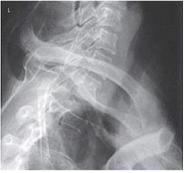
A) MCP at the level of C7-T1
B) MCP at the level of T1-T2
C) MSP at the level of C7-T1
D) MSP at the level of C7 -T1
F) A) and B)
Correct Answer

verified
Correct Answer
verified
Multiple Choice
Where is the central ray directed for a lateral thoracic spine?
A) Level of T5
B) Level of T7
C) Level of T9
D) Level of T10
F) A) and B)
Correct Answer

verified
Correct Answer
verified
Multiple Choice
The intervertebral foramina of the cervical spine are demonstrated on which two of the following projections? (Select all that apply.)
A) AP axial
B) Lateral
C) AP axial oblique
D) PA axial oblique
F) A) and C)
Correct Answer

verified
Correct Answer
verified
Multiple Choice
Which of the following methods is used to evaluate the thoracic and lumbar spine during scoliosis radiography?
A) Pawlow
B) Ferguson
C) Twining
D) Lindblom
F) B) and C)
Correct Answer

verified
Correct Answer
verified
Multiple Choice
An abnormal lateral curvature of the spine is termed:
A) scoliosis.
B) kyphosis.
C) lordosis.
D) scoliokyphosis.
F) A) and B)
Correct Answer

verified
Correct Answer
verified
Multiple Choice
How much is the body rotated for an AP axial oblique projection of the cervical intervertebral foramina?
A) 45 degrees
B) 60 degrees
C) 70 degrees
D) 40 to 60 degrees
F) All of the above
Correct Answer

verified
Correct Answer
verified
Multiple Choice
What anatomy is demonstrated in this AP oblique projection in the image below? 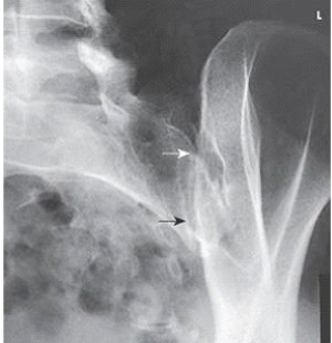
A) Right SI joint
B) Left SI joint
C) L5-S1 joint space
D) L5-S1 zygapophyseal joint
F) A) and D)
Correct Answer

verified
Correct Answer
verified
Multiple Choice
If this is an AP oblique projection,what is the patient position in the image below? 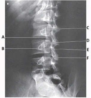
A) 45 degree RPO
B) 45 degree LPO
C) 45 degree RAO
D) 45 degree LAO
F) A) and C)
Correct Answer

verified
Correct Answer
verified
Multiple Choice
The projection demonstrated in this figure is the: 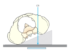
A) AP oblique,sacroiliac joint.
B) PA oblique,sacroiliac joint.
C) AP oblique,lumbar spine.
D) PA oblique,lumbar spine.
F) None of the above
Correct Answer

verified
Correct Answer
verified
Multiple Choice
The respiration phase for the "open mouth" AP projection of the atlas and axis is:
A) inspiration.
B) expiration.
C) suspended respiration.
D) softly phonate "ah" during the exposure.
F) C) and D)
Correct Answer

verified
Correct Answer
verified
Multiple Choice
The articulations between the articular processes of the vertebral arches are called the _____ joints.
A) costovertebral
B) costotransverse
C) intervertebral
D) zygapophyseal
F) C) and D)
Correct Answer

verified
Correct Answer
verified
Multiple Choice
In reference to the ASIS,where is the central-ray entrance for a lateral coccyx?
A) 3 inches (9 cm) posterior and 2 inches (5 cm) inferior
B) 3 inches (9 cm) anterior and 2 inches (5 cm) superior
C) 2 inches (5 cm) posterior and 3 inches (9 cm) inferior
D) 2 inches (5 cm) anterior and 3 inches (9 cm) superior
F) A) and C)
Correct Answer

verified
Correct Answer
verified
Multiple Choice
What anatomy is labeled with the letter B in the image below? 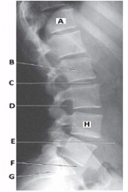
A) Body of L2
B) Body of L3
C) Pedicle of L2
D) Pedicle of L3
F) A) and C)
Correct Answer

verified
Correct Answer
verified
Multiple Choice
The short,thick processes that project posteriorly on each side of a vertebral body are called the:
A) pedicles.
B) laminae.
C) transverse processes.
D) spinous processes.
F) A) and B)
Correct Answer

verified
Correct Answer
verified
Multiple Choice
Where does the central ray enter the body for the AP axial projection (Ferguson method) of the lumbosacral junction?
A) At the pubic symphysis
B) 1 inches superior to the pubic symphysis
C) 3 inches superior to the pubic symphysis
D) At the level of the ASISs
F) C) and D)
Correct Answer

verified
Correct Answer
verified
Multiple Choice
The part identified in this figure is the: 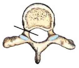
A) vertebral arch.
B) vertebral foramen.
C) intervertebral foramina.
D) transverse foramen.
F) B) and D)
Correct Answer

verified
Correct Answer
verified
Multiple Choice
The part identified in this figure is the: 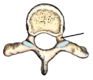
A) body.
B) lamina.
C) pedicle.
D) transverse process.
F) All of the above
Correct Answer

verified
Correct Answer
verified
Multiple Choice
The articulating facet on the superior articular process of the vertebrae is located on its _____ surface.
A) inferior
B) superior
C) anterior
D) posterior
F) None of the above
Correct Answer

verified
Correct Answer
verified
Showing 61 - 80 of 179
Related Exams